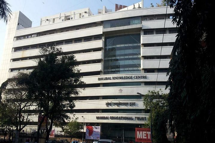
- Medical Hospital
- Library
- Laboratory
- Computer labs
- Auditorium
- Cafeteria
- Sports
Facilities :-
Library
The Library moved into its new location on the 6 th floor of the existing building in August 2006. The new and improved library is located in 2500 sq ft space provided on the top floor of the building. The library is fully airconditioned and has a superb aerial view of the vicinity from its high vantage point owing to windows along its three sides. Its location provides for an ideal place for readers to come and collect their thoughts or read or browse on the internet. Books and journals are housed in compactors (movable racks). Additional storage space has been provided along the walls in such a way so as not to disturb the aesthetics of the building. Readers' consoles provide the seclusion needed for concentrated reading both for scientists and students.
The library presently subscribes 120 national and international journals and has an excellent collection of books, monographs, atlases etc. in the areas of biochemistry, immunology, endocrinology, stem cell biology, structural biology and bioinformatics.
Transmission Electron Microscopy : Central Core facility
The Transmission Electron Microscope (TEM) allows the user to determine the internal structure of materials, either of biological or non-biological origin to reveal the finest details of internal structure - in some cases as small as individual atoms. For biological samples, cell structure and morphology is commonly determined whilst the localization of antigens or other specific components within cells is readily undertaken using specialized preparative techniques.
The role of the TEM facility at NIRRH is to promote, support and initiate research and teaching in the applications of electron microscopy. The TEM at the institute has a resolution of 0.2nm at 100kV. Sub-cellular details are easily envisaged at magnifications of about 3x10⁵. The facility is accessible to the scientists and students who require cellular biology-oriented high-resolution imaging.
The facility undertakes all the steps required for TEM including sample preparation, analysis at semithin levels followed by ultrathin sectioning and imaging. Users are expected to get the fixed samples in desired fixatives and participate actively in scanning and data analysis.
Confocal Laser Scanning Microscope
Confocal microscopy offers several advantages over conventional optical microscopy, including controllable depth of field, the elimination of image degrading out-of-focus information, and the ability to collect serial optical sections from thick specimens.
The Institute has a Confocal Laser Scanning Microscope (LSM) with spectral imaging. The system is mounted on an inverted microscope with temperature controlled stage. The laser systems include the Argon and Helium-neon lasers for fluorescence excitation of 458/488, 477, 514 and 633 nm, the DPSS and blue diode laser for 561, 405 nm excitation wavelengths. The scanning module has two single-channel detectors and polychromatic multichannel detector capable of simultaneous acquisition of up to 8 channels. Beside the feature of high resolution optical slicing and 3 dimensional imaging of the cells and tissues, it can also perform FRAP, FRET, FLIM and live cell imaging studies.
Investigators perform the labeling/staining as per requirements using appropriate flourophores in their laboratory. The system is available for viewing and analysis. Presently we have the expertise in co-localization and 3D reconstruction of the samples. Future plans are to include FRET and FRAP analysis as well.
Microarray
Analysis of gene expression has become an integrated approach towards understanding the biological system, drug effects, diseases etc, However, in most instances comparisons are done in a limited number of genes. But for meaningful interpretations a large number of genes need to be analyzed. Microarray is a cutting edge technology that permits analysis of several thousand genes simultaneously. Using this technology it is now possible to obtain gene patterns of several tissues and diseases. In fact it is now possible to diagnose diseases based on microarray technology.
The Microarray Core Facility is established to provide the infrastructure for the development of major projects that utilize DNA microarrays for large scale analysis of gene expression. The facility provides researchers to hybridize and read prespotted microarrays from most sources. Investigators can bring their labeled samples and slides for hybridization, scanning and data analysis. The facility also has linkages with the Medical Informatics Center at NIRRH for data mining using academic tools.
The facility further aims at developing protocols for other microarray based applications like SNP genotyping, CGH arrays antibody arrays, protein arrays and tissue arrays.
Laser Capture Microdissection
The laser capture microdissection permits procurement of DNA, RNA and proteins of small and pure populations of cells. Traditionally used for separation of tumor cells, many additional applications of this technology, e.g. for developmental biology, or in vitro fertilization have evolved. In addition, the combination of laser capture microscopy with other high sensitivity, high-throughput technologies such as microarrays offers exciting new possibilities for scientists in research and clinic.
The Institute has a Laser Capture Microdissection Microscope featuring a brightfield upright microscope with fluorescent and phase attachment. It is used to procure pure population of cells from microscopic tissue sections. The facility provides staining and microdissection assistance to the investigators.
Flow Cytometer Core facility
Flow Cytometry is a powerful tool which can simultaneously measure multiple characteristics of single cell such as cell surface and intracellular antigen expression, light scattering properties, DNA ploidy, cell cycle analysis, apoptosis at a rapid rate.
The facility is equipped with a state-of-the-art flow cytometry and multiparameter cell sorting instrumentation with four laser system (355, 488, 532 and 633nm) capable of fifteen parameter analysis and cell sorting. Its acquisition software is FACSDiva 8. The facility permits maximum and efficient utilization of the equipment and serves to enhance the quality and scope of scientific research performed at NIRRH. The core facility provides instrumentation, technical and professional assistance for flow cytometry experiments.
DNA Sequencing Facility
The Institute has set up a core facility for DNA sequencing mainly to support the projects. The facility offers high-quality capillary-based DNA sequencing. The facility is outfitted with two equipments that include a 4 capillary and a 16 capillary sequencer. Along with standard DNA sequencing, the setup also offers Fragment Analysis for microsatellites, AFLP, SNP's or any other fragment applications.
Users provide DNA (plasmid, phage or PCR product) at a standardized concentration and custom primers if necessary. The Facility performs cycle sequencing reactions using fluorescent dye terminators, carries out electrophoresis, acquires the data, and provides the sequence as chromatogram and text files.
Proteomics Workstation
Proteomics is the study and identification of the thousands of types of proteins found in humans, plants, animals, bacteria and other life forms, and the expression of particular proteins can be used as “biomarkers” of health, disease and/or quality.
The newly established Proteomics Facility within the Institute allows users to isolate and identify proteins of interest. The proteomics workstation includes gel documentation system, DIGE scanner, 2D analysis software's, automated spot picker, spot digester and MALDI target spotter. We use the MALDI TOF-TOF based mass spectrometry for molecular weight determination and derivation of sequence tags of proteotically cleaved peptides.
The investigators are expected to run and analyze their own 2D gels. In consultation with the staff of the facility they can provide gels or manually cored spots (Silver, Comassie or fluorescently stained) for further analysis. The facility offers automated coring of spots, tryptic digestion and analysis of peptides using mass spectrometry. Preliminary bioinformatics based analysis is also provided for protein identification. Investigators can avail the Medial Informatics Core facility for further in silico analysis.
Peptide Synthesis Facility
The Institute has a specialized facility for the chemical synthesis of peptides of desired amino acid sequences. The assembled peptide is cleaved from the solid support using appropriate cleavage cocktails, purified using preparative RP-HPLC and characterized by amino acid analysis and MALDI-TOF. The peptides are assembled using solid phase technology and Fmoc-chemistry.
The users are expected to provide the sequence of the peptides (up to 20 residues max) that need to be synthesized. The purified synthetic peptides will be provided in dried form along with the RP-HPLC profiles, MALDI-TOF spectra and amino acid composition of the synthesized peptide.
Amino Acid Analysis Facility
The Institute has a facility to perform the amino acid analysis of any peptide/ protein of interest. The peptide/protein is hydrolyzed followed by ion exchange chromatography using a dedicated amino acid analyzer. This provides identification of the constituent amino acids present in the peptide/protein. The resultant chromatogram gives the identity, ratio and amount of the amino acids present in the given sample.
The users have to provide pure peptide or protein in the concentration of ~ 500 nmoles for analysis.
Circular Dichroism Spectroscopy Facility
The Institute has the facility for recording the circular dichroism spectra of peptide/protein molecules using the spectropolarimeter. The instrument software converts the CD signals into secondary structure information i.e. alpha helix, beta sheet, beta turn or random coil of polypeptides and proteins for analysis.
The users need to provide micromolar concentrations of pure peptide or protein for spectral analysis. The users will be provided with the spectra generated by the system and the computer based analysis of the output.
Real-Time PCR
Real-Time PCR or Q-PCR has now become an essential component of studies based on quantitation of gene expression, array verification, pathogen detection, viral load estimation and genotyping. In contrast to traditional PCR methods, Real-Time PCR allows measuring the kinetics of the amplification reaction in the initial phases. Traditional methods use time-consuming gel electrophoresis for detection of PCR amplification only at the end of the PCR reaction. Further these methods have limitations such as poor precision, low sensitivity, short dynamic range, low resolution, non-automation etc. In contrast, Real time PCR collects data in the exponential growth phase and quantifies the number of amplicons on the basis of fluorescent signals generated. Further Real Time PCR offers increased dynamic range of detection without any post PCR processing of the amplified samples.
Investigators at the institute are using this facility for quantifying the expression levels of various genes in reproductive tissues/organs under different physiological or pathological conditions. The facility is also being used for SNP genotyping of individuals predisposed to certain reproductive disorders such as congenital adrenal hyperplasia etc. Users bring their Q-PCR reaction samples in a system-compatible (Applied Biosystems-7900HT) 96-well plate format with optical adhesive covers. The system provides data for relative/absolute quantification, melt curve and allele discrimination.
Experimental Animal Facility
The animal facility is distributed over three floors with an area of nearly 8000 sq ft. and is supported with qualified veterinarians and skilled animal care staff. The facility meets the requirements as per the guidelines of Committee for the Purpose of Control & Supervision of Experiments on Animals ( CPCSEA) and is registered with the CPCSEA. An Institutional Animal Ethics Committee (IAEC) has been constituted as per CPCSEA guidelines and reviews the scientific projects of the institute quarterly.
The facility undertakes animal breeding, management, care and husbandry practices. The facility houses different laboratory animals viz. mice, rats, rabbits and nonhuman primates like marmosets and bonnet monkeys. The facility is engaged in the production of quality animals by undertaking various quality control programs and preventive measures. The facility has Individual Ventilated Caging System (IVC) to house rodents to prevent cross-contamination.
